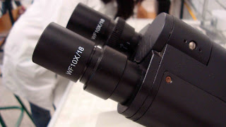
En el informe que un alumno estaba escribiendo en el laboratorio hay un resultado incorrecto, una respuesta parcialmente correcta y falta un resultado. Además hay un error ortográfico imperdonable... Si el próximo martes me dices cuáles eran los errores y las respuestas correctas... serás recompenzado generosamente con algunas décimas de punto... en la calificación bimestral...(también puedes editar un comentario en este blog con tus respuestas)
PULGAS!!!... de un gato: El material biológico fué aportado por una estudiante de la segunda sección de laboratorio y ... ganó el punto bimestral... Gracias y felicidades por tu colaboración y entusiasmo demostrado durante la observación al microscopio...
Pulga vista a 40 x
imagen a 100x
 | |
| Ciclo de vida de las pulgas |
Para determinar el aumento al que estás viendo las imágenes al microscopio óptico, recuerda que debes multiplicar el aumento que te proporciona el ocular (en este caso 10 x ) ...
... por el aumento que te brinda el objetivo... que en el caso ilustrado es de 4 x... por lo tanto esa combinación de lentes te brindaría un aumento de _____ (puedes dar la respuesta editando un comentario en este blog)
Y para saber más sobre el tema te recomiendo consultar la obra de Robert Hooke, quién en 1665 publicó su obra maestra MICROGRAPHIA, y en ella dibujo una pulga:
 | ||||||||||||||
| Para ver una versión digital del libro original, da clik aquí |
Y de la página de la Universidad de Berkeley tomamos la siguiente información:
Robert Hooke (1635-1703)
Hooke's reputation in the history of biology largely rests on his book Micrographia, published in 1665. Hooke devised the compound microscope and illumination system shown above, one of the best such microscopes of his time, and used it in his demonstrations at the Royal Society's meetings. With it he observed organisms as diverse as insects, sponges, bryozoans, foraminifera, and bird feathers. Micrographia was an accurate and detailed record of his observations, illustrated with magnificent drawings, such as the flea shown below, which Hooke described as "adorn'd with a curiously polish'd suite of sable Armour, neatly jointed. . ." It was a best-seller of its day. Some readers ridiculed Hooke for paying attention to such trifling pursuits: a satirist of the time poked fun at him as "a Sot, that has spent 2000 £ in Microscopes, to find out the nature of Eels in Vinegar, Mites in Cheese, and the Blue of Plums which he has subtly found out to be living creatures." More complimentary was the reaction of the diarist and government official Samuel Pepys, who stayed up till 2:00 AM one night reading Micrographia, which he called "the most ingenious book that I ever read in my life."
Perhaps his most famous microscopical observation was his study of thin slices of cork, depicted above right. In "Observation XVIII" of the Micrographia, he wrote:
. . . I could exceedingly plainly perceive it to be all perforated and porous, much like a Honey-comb, but that the pores of it were not regular. . . . these pores, or cells, . . . were indeed the first microscopical pores I ever saw, and perhaps, that were ever seen, for I had not met with any Writer or Person, that had made any mention of them before this. . .
Hooke had discovered plant cells -- more precisely, what Hooke saw were the cell walls in cork tissue. In fact, it was Hooke who coined the term "cells": the boxlike cells of cork reminded him of the cells of a monastery. Hooke also reported seeing similar structures in wood and in other plants. In 1678, after Leeuwenhoek had written to the Royal Society with a report of discovering "little animals" -- bacteria and protozoa -- Hooke was asked by the Society to confirm Leeuwenhoek's findings. He successfully did so, thus paving the way for the wide acceptance of Leeuwenhoek's discoveries. Hooke noted that Leeuwenhoek's simple microscopes gave clearer images than his compound microscope, but found simple microscopes difficult to use: he called them "offensive to my eye" and complained that they "much strained and weakened the sight."
 |
| Lámina XI del libro Micrographia que muestra las estructuras que Hooke describió como células. Hoy sabemos que corresponden a las paredes celulares que dejaron las células que formaron el tejido del corcho. |


no encuentro la séptima sesión
ResponderEliminarLos comentarios a entradas del blog aparecen como si yo los dijera, debido a que se ingresa con la cuenta de correo con la que estoy idcentificado... intenta hacerlo desde la tuya... Para empezar ¿quién eres?... y en segundo lugar... ¿que te te parece si tu escribes una nota sobre esa sesión?...atte: Julio
ResponderEliminar